21+ Internal Anatomy Of The Kidney Including The Nephron
21+ Internal Anatomy Of The Kidney Including The Nephron. By anatomical studies using light the distal convoluted tubule is the nephron segment that lies immediately downstream of the macula densa. It surrounds a tuft of capillaries called the glomerulus that carries blood from the renal arteries into the nephron, where plasma is filtered.
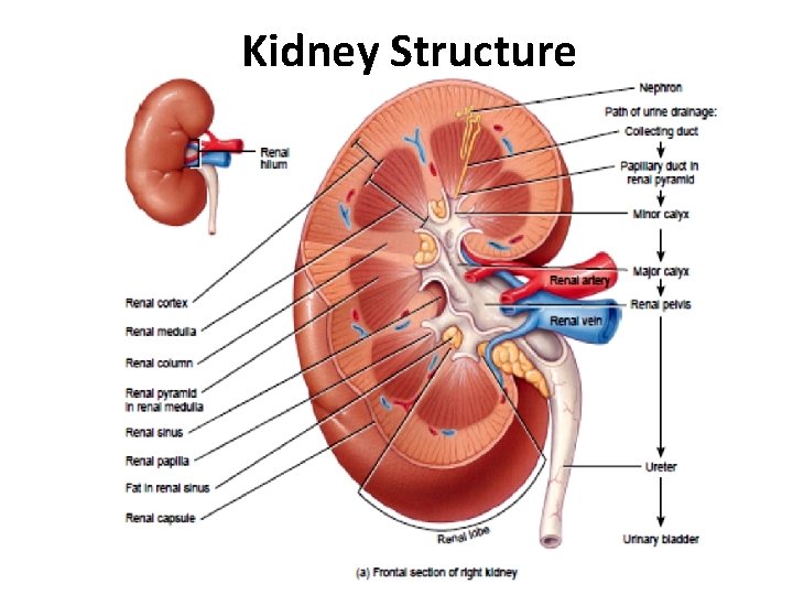
Internally, kidneys are mainly composed of over one million nephrons and an extensive network of blood vessels and capillaries.
The kidneys are important in maintaining the internal environment: The functional part of the kidneys that filters the blood (renal corpuscle), reabsorbs minerals/water and secretes waste (renal tubule), and produces the substance called urine which will drain down into the. The kidneys have a tough organ capsule (capsula fibrosa), which follows gerota's fascia surrounds the kidney, including perinephric fat and the adrenal gland. Nephrons extend through the cortex and medulla areas of the.


Comments
Post a Comment