14+ Anatomy Of Heart And Blood Vessels. Together, the heart, blood, and blood vessels — arteries, capillaries, and veins — make up the circulatory system. The heart is a muscular organ that pumps blood around the body by circulating it through the circulatory/vascular system.
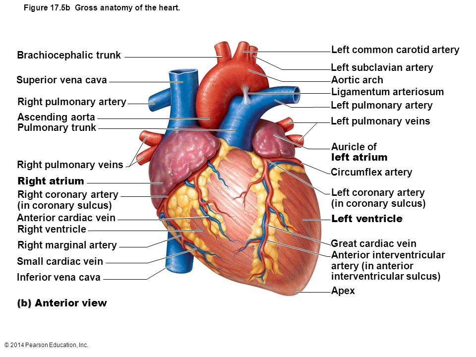
Figure 17 5b Gross Anatomy Of The Heart Ppt Video Online Download from slideplayer.com All blood vessels are specifically structured to perform their function. Blood vessels are intricate networks of hollow tubes that transport blood throughout the entire body. The heart is tilted so that its pointed end, the apex, points downward toward the left hi.
Veins are the large blood vessels that return blood from various tissues to the heart so that the blood can be pumped out to tissues again.
Intraoperatively, the anatomy of the heart is viewed from the right side of the supine patient via a median sternotomy incision. The heart has its own set of blood vessels that provide the myocardium with the oxygen and nutrients necessary to pump blood throughout the body. Carbon dioxide exits the blood and oxygen enters. Atria are often called the receiving chambers because they receive blood from vessels called veins.
42+ Ct Anatomy Of Neck Spaces . Posted by radiologypics ⋅ march 21, 2013 ⋅ 1 comment. Atlas of the anatomy of the head and neck on a ct in axial, coronal, and sagittal sections, and 3d images. Neck Spaces 1 Parapharyngeal Space 2 Masticator Space 3 Carotid Space 4 Parotid Space 5 Mucosal Space 6 Perivertebral Radiology Imaging Radiology Anatomy from i.pinimg.com The 5 anatomical spaces of the infrahyoid neck. They are only created by pathology, e.g. 875 x 507 jpeg 87 кб. The anatomy of the neck is complex and, as a result, not very well understood. Deep neck space infections michael d. Ct anatomy of neck spaces rv. Learn vocabulary, terms and more with flashcards, games and other study tools. Fasciae & spaces of the neck. Source: pubs.rsna.org They are only created by pathology, e.g. Source: i2.wp.com Neck nod...
11+ Human Anatomy Abdomen Muscles . Next to it on both sides of the body is the internal oblique. The posterior abdominal wall is found medial to the. Core Stability Abdominals Back Pain Pouch Weak Core Priority Physical Therapy Llc from images.squarespace-cdn.com An interactive demonstration of the transversus abdominis muscle (insertion, origin, actions & innervations) featuring the iconic gbs illustrations. You can locate them by putting your the external oblique muscles allow flexion of the spine, rotation of the torso, sideways bending and compression of the abdomen. The posterior abdominal wall is found medial to the. Actions.—when the pelvis and thorax are fixed, the abdominal muscles compress the abdominal viscera by constricting the cavity of the abdomen, in which action they are materially assisted by the. Attached to the bones of the skeletal abdomen and lower back. Att...
28+ Skull Base Ct Anatomy . 2.1 skull base anatomy—anterior and middle surgical anatomy pearl. The bregma is the point where joins coronal suture and sagittal suture. Middle Skull Base Plastic Surgery Key from plasticsurgerykey.com The skull is a bony structure that supports the face and forms a protective cavity for the brain. This view may expose some daunting anatomy, but it is not difficult to learn once you associate the various structures with a function and in that way store all the informations into your. 2.1 skull base anatomy—anterior and middle surgical anatomy pearl. The sphenoid bone is a single, complex bone of the central skull (). Skull base anatomy and cranial nerves explained in a simple way to remember it and use it in your daily practice as a radiation. It shows the thin cortical margins of skull base neurovascular foramina. Inferior view of base of the skull. The ...


Comments
Post a Comment