28+ Anatomy Of Intestines. They are connected to the posterior wall of the abdomen by the mesentery, a thin vascular membrane. The small intestine is a organ located in the gastrointestinal tract, which assists in the digestion and absorption of ingested food.
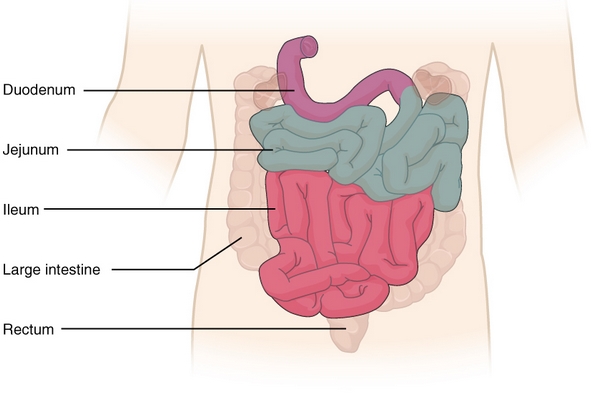
Small Intestine Pathology Structure Duodenum Jejunum Ileum from d3uigcfkiiww0g.cloudfront.net The gi tract has a similar layout through out its length: Satheesha nayak b, professor at department of anatomy of melaka manipal medical college mmmc (mahe), manipal. The intestines absorb nutrients and vitamins and are part of the gastrointestinal (gi) tract.
Lymph vessels and nodes of large intestine anatomy, paracolic nodes, middle colic nodes, superior mesenteric nodes (central superior group), right colic nodes, ileocolic nodes, prececal nodes.
Anatomy and histology of small intestine simple explaination in hindi | bhushan science in this video, some of the details of anatomy of large intestine have been described. The intestines are located inferior to the stomach in the abdominal body cavity. Online quizzes for cliffsnotes anatomy and physiology quickreview, 2nd edition. We only think about our intestines when we have intestinal pain, or when something else goes the intestines begin with the small intestine, divided into three parts whimsically named the duodenum.
42+ Ct Anatomy Of Neck Spaces . Posted by radiologypics ⋅ march 21, 2013 ⋅ 1 comment. Atlas of the anatomy of the head and neck on a ct in axial, coronal, and sagittal sections, and 3d images. Neck Spaces 1 Parapharyngeal Space 2 Masticator Space 3 Carotid Space 4 Parotid Space 5 Mucosal Space 6 Perivertebral Radiology Imaging Radiology Anatomy from i.pinimg.com The 5 anatomical spaces of the infrahyoid neck. They are only created by pathology, e.g. 875 x 507 jpeg 87 кб. The anatomy of the neck is complex and, as a result, not very well understood. Deep neck space infections michael d. Ct anatomy of neck spaces rv. Learn vocabulary, terms and more with flashcards, games and other study tools. Fasciae & spaces of the neck. Source: pubs.rsna.org They are only created by pathology, e.g. Source: i2.wp.com Neck nod...
11+ Human Anatomy Abdomen Muscles . Next to it on both sides of the body is the internal oblique. The posterior abdominal wall is found medial to the. Core Stability Abdominals Back Pain Pouch Weak Core Priority Physical Therapy Llc from images.squarespace-cdn.com An interactive demonstration of the transversus abdominis muscle (insertion, origin, actions & innervations) featuring the iconic gbs illustrations. You can locate them by putting your the external oblique muscles allow flexion of the spine, rotation of the torso, sideways bending and compression of the abdomen. The posterior abdominal wall is found medial to the. Actions.—when the pelvis and thorax are fixed, the abdominal muscles compress the abdominal viscera by constricting the cavity of the abdomen, in which action they are materially assisted by the. Attached to the bones of the skeletal abdomen and lower back. Att...
28+ Skull Base Ct Anatomy . 2.1 skull base anatomy—anterior and middle surgical anatomy pearl. The bregma is the point where joins coronal suture and sagittal suture. Middle Skull Base Plastic Surgery Key from plasticsurgerykey.com The skull is a bony structure that supports the face and forms a protective cavity for the brain. This view may expose some daunting anatomy, but it is not difficult to learn once you associate the various structures with a function and in that way store all the informations into your. 2.1 skull base anatomy—anterior and middle surgical anatomy pearl. The sphenoid bone is a single, complex bone of the central skull (). Skull base anatomy and cranial nerves explained in a simple way to remember it and use it in your daily practice as a radiation. It shows the thin cortical margins of skull base neurovascular foramina. Inferior view of base of the skull. The ...


Comments
Post a Comment