47+ Labeled Internal Anatomy Of Kidney. Urology addresses diseases of kidney (and urinary tract) anatomy: Its parts are labelled except the (gray) the first to examine the ureter through an internal approach, called ureteroscopy, rather than surgery.
Kidney Diagram Nephron Stock Illustrations 101 Kidney Diagram Nephron Stock Illustrations Vectors Clipart Dreamstime from thumbs.dreamstime.com The kidneys are a paired organ; The kidneys are made up by three external layers, which include the renal fascia (the outermost layer), the perirenal fat capsule, and lastly, the innermost layer, the renal capsule, which then surround the space. Bacteria may infect the kidney, usually causing back pain and fever.
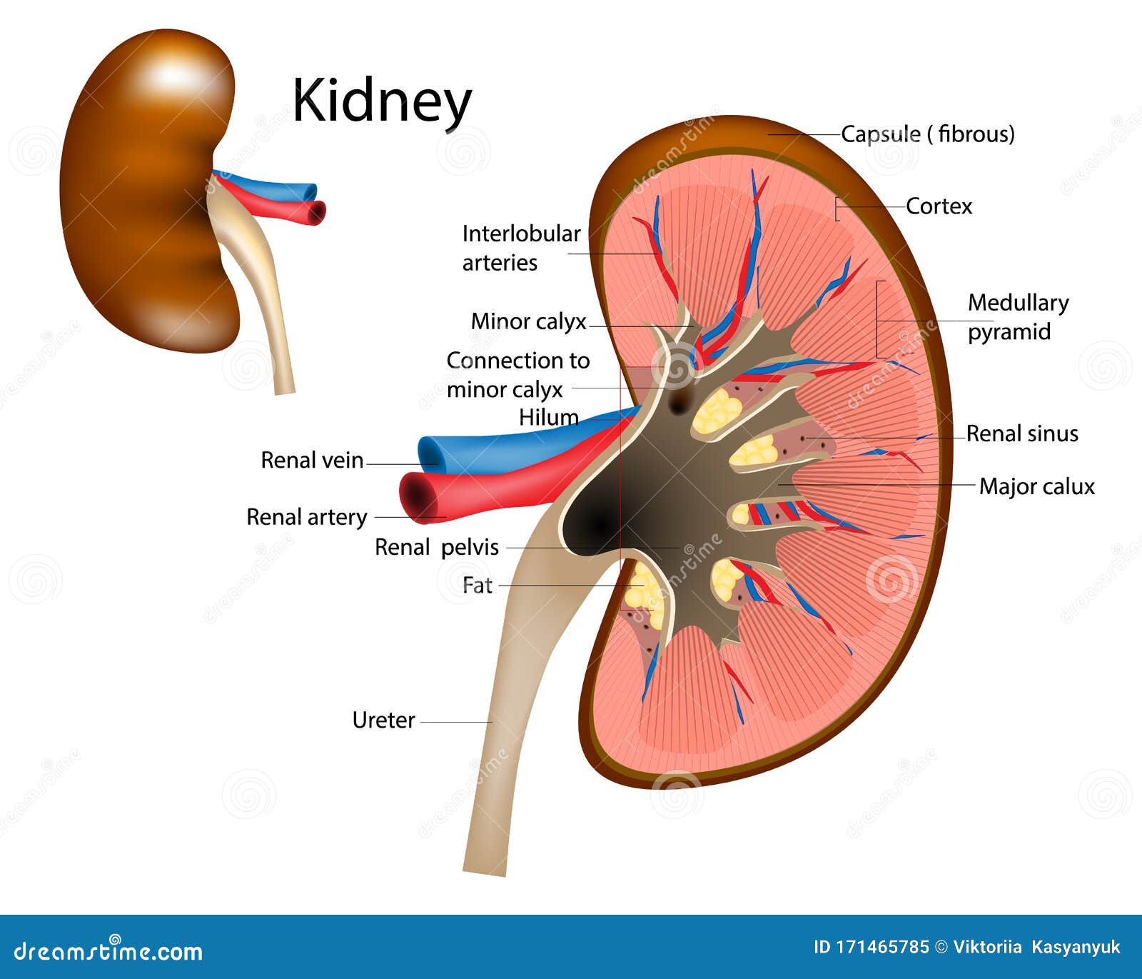
The kidneys are a pair of organs found along the posterior muscular wall of the abdominal cavity.
Its parts are labelled except the (gray) the first to examine the ureter through an internal approach, called ureteroscopy, rather than surgery. Kidneys anatomy function and internal. Each kidney is enclosed by a thin tough fibrous connective tissue called renal capsule that protects it from infections and injuries. Gross anatomy location the kidneys are located on the posterior abdominal wall, with one on either side of the vertebral column, in the peri.
42+ Ct Anatomy Of Neck Spaces . Posted by radiologypics ⋅ march 21, 2013 ⋅ 1 comment. Atlas of the anatomy of the head and neck on a ct in axial, coronal, and sagittal sections, and 3d images. Neck Spaces 1 Parapharyngeal Space 2 Masticator Space 3 Carotid Space 4 Parotid Space 5 Mucosal Space 6 Perivertebral Radiology Imaging Radiology Anatomy from i.pinimg.com The 5 anatomical spaces of the infrahyoid neck. They are only created by pathology, e.g. 875 x 507 jpeg 87 кб. The anatomy of the neck is complex and, as a result, not very well understood. Deep neck space infections michael d. Ct anatomy of neck spaces rv. Learn vocabulary, terms and more with flashcards, games and other study tools. Fasciae & spaces of the neck. Source: pubs.rsna.org They are only created by pathology, e.g. Source: i2.wp.com Neck nod...
11+ Human Anatomy Abdomen Muscles . Next to it on both sides of the body is the internal oblique. The posterior abdominal wall is found medial to the. Core Stability Abdominals Back Pain Pouch Weak Core Priority Physical Therapy Llc from images.squarespace-cdn.com An interactive demonstration of the transversus abdominis muscle (insertion, origin, actions & innervations) featuring the iconic gbs illustrations. You can locate them by putting your the external oblique muscles allow flexion of the spine, rotation of the torso, sideways bending and compression of the abdomen. The posterior abdominal wall is found medial to the. Actions.—when the pelvis and thorax are fixed, the abdominal muscles compress the abdominal viscera by constricting the cavity of the abdomen, in which action they are materially assisted by the. Attached to the bones of the skeletal abdomen and lower back. Att...
28+ Skull Base Ct Anatomy . 2.1 skull base anatomy—anterior and middle surgical anatomy pearl. The bregma is the point where joins coronal suture and sagittal suture. Middle Skull Base Plastic Surgery Key from plasticsurgerykey.com The skull is a bony structure that supports the face and forms a protective cavity for the brain. This view may expose some daunting anatomy, but it is not difficult to learn once you associate the various structures with a function and in that way store all the informations into your. 2.1 skull base anatomy—anterior and middle surgical anatomy pearl. The sphenoid bone is a single, complex bone of the central skull (). Skull base anatomy and cranial nerves explained in a simple way to remember it and use it in your daily practice as a radiation. It shows the thin cortical margins of skull base neurovascular foramina. Inferior view of base of the skull. The ...


Comments
Post a Comment