48+ Femoral Nerve And Artery Anatomy. The femoral nerve is a nerve in the thigh that supplies skin on the upper thigh and inner leg, and the muscles that extend the knee. Femoral nerve is the major nerve supplying the anterior compartment of the thigh.
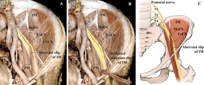
Femoral Nerve Split With Variant Iliacus Muscle A Potential Source Of Femoral Nerve Entrapment Springerlink from media.springernature.com Profunda femoris arises on the lateral side of the femoral artery in the femoral triangle about 1.5 inch (4 cm) below the inguinal ligament. It originates from the dorsal sections of the anterior primary rami of l2, l3, l4 nerves and is the largest branch the lateral circumflex femoral artery is straddled by both sections. Lower abdomen and perineal region • superficial epigastric artery • superficial circumflex iliac artery • superficial external pudendal artery deep branches • profunda femoris artery • main branch of the femoral.
It exits the pelvis through the sciatic notch, and continues down the back of the thigh where it splits into the common fibular and tibial nerve at the knee.
Femoral nerve is the main nerve of anterior compartment of thigh. The artery is superficial to the adductor magnus and longus muscles. Its muscular branches innervate the four quadriceps muscles (rectus femoris, vastus lateralis, vastus medialis, and vastus intermedius), which flex the thigh at the hip and extend. It crosses under the inguinal ligament and enters the femoral triangle where the profunda femoris artery arises from its lateral side.
42+ Ct Anatomy Of Neck Spaces . Posted by radiologypics ⋅ march 21, 2013 ⋅ 1 comment. Atlas of the anatomy of the head and neck on a ct in axial, coronal, and sagittal sections, and 3d images. Neck Spaces 1 Parapharyngeal Space 2 Masticator Space 3 Carotid Space 4 Parotid Space 5 Mucosal Space 6 Perivertebral Radiology Imaging Radiology Anatomy from i.pinimg.com The 5 anatomical spaces of the infrahyoid neck. They are only created by pathology, e.g. 875 x 507 jpeg 87 кб. The anatomy of the neck is complex and, as a result, not very well understood. Deep neck space infections michael d. Ct anatomy of neck spaces rv. Learn vocabulary, terms and more with flashcards, games and other study tools. Fasciae & spaces of the neck. Source: pubs.rsna.org They are only created by pathology, e.g. Source: i2.wp.com Neck nod...
11+ Human Anatomy Abdomen Muscles . Next to it on both sides of the body is the internal oblique. The posterior abdominal wall is found medial to the. Core Stability Abdominals Back Pain Pouch Weak Core Priority Physical Therapy Llc from images.squarespace-cdn.com An interactive demonstration of the transversus abdominis muscle (insertion, origin, actions & innervations) featuring the iconic gbs illustrations. You can locate them by putting your the external oblique muscles allow flexion of the spine, rotation of the torso, sideways bending and compression of the abdomen. The posterior abdominal wall is found medial to the. Actions.—when the pelvis and thorax are fixed, the abdominal muscles compress the abdominal viscera by constricting the cavity of the abdomen, in which action they are materially assisted by the. Attached to the bones of the skeletal abdomen and lower back. Att...
28+ Skull Base Ct Anatomy . 2.1 skull base anatomy—anterior and middle surgical anatomy pearl. The bregma is the point where joins coronal suture and sagittal suture. Middle Skull Base Plastic Surgery Key from plasticsurgerykey.com The skull is a bony structure that supports the face and forms a protective cavity for the brain. This view may expose some daunting anatomy, but it is not difficult to learn once you associate the various structures with a function and in that way store all the informations into your. 2.1 skull base anatomy—anterior and middle surgical anatomy pearl. The sphenoid bone is a single, complex bone of the central skull (). Skull base anatomy and cranial nerves explained in a simple way to remember it and use it in your daily practice as a radiation. It shows the thin cortical margins of skull base neurovascular foramina. Inferior view of base of the skull. The ...


Comments
Post a Comment