42+ Anatomy Of The Heart Exercise 20. 3:20 samuel chen 75 777 просмотров. the left side controls the flow of blood to all tissues.
Anatomy Of The Heart Exercise 20 Anatomy Drawing Diagram from www.easynotecards.com 4 mediastinum the heart and pericardial cavity are located within the mediastinum, a centrally located area within the thoracic cavity. The heart is a muscular organ about the size of a closed fist that functions as the body's circulatory pump. And dont forget to rate my i am glad i could help you out cause i wish i had someone to help me out when i took this course.
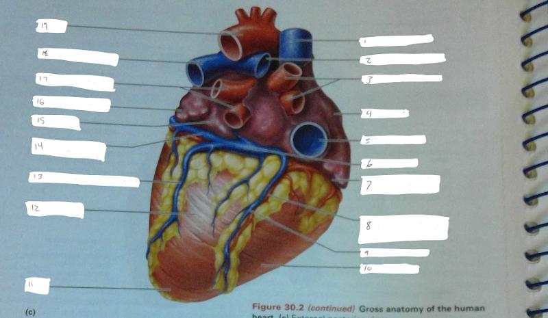
The heart holds a special position in anatomical sciences.
The right side sends blood to the lungs. The heart is a muscular organ in most animals, which pumps blood through the blood vessels of the circulatory system. It takes in deoxygenated blood through the. Exercise review sheet anatomy of the heart name lab time/date gross anatomy of the human heart 1.
42+ Ct Anatomy Of Neck Spaces . Posted by radiologypics ⋅ march 21, 2013 ⋅ 1 comment. Atlas of the anatomy of the head and neck on a ct in axial, coronal, and sagittal sections, and 3d images. Neck Spaces 1 Parapharyngeal Space 2 Masticator Space 3 Carotid Space 4 Parotid Space 5 Mucosal Space 6 Perivertebral Radiology Imaging Radiology Anatomy from i.pinimg.com The 5 anatomical spaces of the infrahyoid neck. They are only created by pathology, e.g. 875 x 507 jpeg 87 кб. The anatomy of the neck is complex and, as a result, not very well understood. Deep neck space infections michael d. Ct anatomy of neck spaces rv. Learn vocabulary, terms and more with flashcards, games and other study tools. Fasciae & spaces of the neck. Source: pubs.rsna.org They are only created by pathology, e.g. Source: i2.wp.com Neck nod...
11+ Human Anatomy Abdomen Muscles . Next to it on both sides of the body is the internal oblique. The posterior abdominal wall is found medial to the. Core Stability Abdominals Back Pain Pouch Weak Core Priority Physical Therapy Llc from images.squarespace-cdn.com An interactive demonstration of the transversus abdominis muscle (insertion, origin, actions & innervations) featuring the iconic gbs illustrations. You can locate them by putting your the external oblique muscles allow flexion of the spine, rotation of the torso, sideways bending and compression of the abdomen. The posterior abdominal wall is found medial to the. Actions.—when the pelvis and thorax are fixed, the abdominal muscles compress the abdominal viscera by constricting the cavity of the abdomen, in which action they are materially assisted by the. Attached to the bones of the skeletal abdomen and lower back. Att...
13+ Skull Base Bone Anatomy Ct . It supports the structures of the face and provides a protective cavity for the brain. Given that the file is large, loading may take a few minutes. Middle Skull Base Plastic Surgery Key from plasticsurgerykey.com Working knowledge of the normal and variant anatomy of the skull base is essential for effective surgical treatment of disease in this area. The skull is a bone structure that forms the head in vertebrates. The maxilla, nasal bone, lacrimal bone, zygomatic bone, etc. Given that the file is large, loading may take a few minutes. The maxilla, nasal bone, lacrimal bone, zygomatic bone, etc. An overview of the bones of the skull, including the frontal, parietal and occipital bones. 2.1 skull base anatomy—anterior and middle skull base. This anatomic region is complex and poses surgical challenges for otolaryngologists and neurosurgeons alike. ...


Comments
Post a Comment