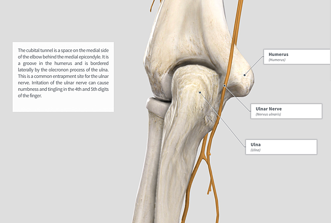
35+ Ulnar Groove Anatomy. Original editor trevor bradshaw ; Groove for ulnar nerve is a groove on the back of the medial epicondyle where the ulnar nerve runs.
Cubital Tunnel Anatomy Anatomy Drawing Diagram from images.squarespace-cdn.com The transformation of the groove into a tunnel takes place, which is called cubital tunnel by a fibrous band extending between medial epicondyle and. Into a bony groove on humerus (cubital tunnel). Groove for ulnar nerve is a groove on the back of the medial epicondyle where the ulnar nerve runs.
The ulnar groove does not appear on the dorsal side of the medial humeral epicondyle earlier than an aponeurotic arch bridging the humerus and ulna and covering the ulnar nerve medially could not.
The humerus along the spiral groove.the lower portion of the radial nerve crosses the midline at an average of15 cm from the. Anatomy of ulnar nerve (ulnar nerve anatomy). The ulnar groove is shallow and the nerve may become more prominent than the medial epicondyle or the loss of the ulnar groove may be associated with arthritis of the elbow joint, often due to an old. Groove anatomy are the ultimate function/party band comprising six young, versatile and extremely talented musicians.
42+ Ct Anatomy Of Neck Spaces . Posted by radiologypics ⋅ march 21, 2013 ⋅ 1 comment. Atlas of the anatomy of the head and neck on a ct in axial, coronal, and sagittal sections, and 3d images. Neck Spaces 1 Parapharyngeal Space 2 Masticator Space 3 Carotid Space 4 Parotid Space 5 Mucosal Space 6 Perivertebral Radiology Imaging Radiology Anatomy from i.pinimg.com The 5 anatomical spaces of the infrahyoid neck. They are only created by pathology, e.g. 875 x 507 jpeg 87 кб. The anatomy of the neck is complex and, as a result, not very well understood. Deep neck space infections michael d. Ct anatomy of neck spaces rv. Learn vocabulary, terms and more with flashcards, games and other study tools. Fasciae & spaces of the neck. Source: pubs.rsna.org They are only created by pathology, e.g. Source: i2.wp.com Neck nod...
11+ Human Anatomy Abdomen Muscles . Next to it on both sides of the body is the internal oblique. The posterior abdominal wall is found medial to the. Core Stability Abdominals Back Pain Pouch Weak Core Priority Physical Therapy Llc from images.squarespace-cdn.com An interactive demonstration of the transversus abdominis muscle (insertion, origin, actions & innervations) featuring the iconic gbs illustrations. You can locate them by putting your the external oblique muscles allow flexion of the spine, rotation of the torso, sideways bending and compression of the abdomen. The posterior abdominal wall is found medial to the. Actions.—when the pelvis and thorax are fixed, the abdominal muscles compress the abdominal viscera by constricting the cavity of the abdomen, in which action they are materially assisted by the. Attached to the bones of the skeletal abdomen and lower back. Att...
13+ Skull Base Bone Anatomy Ct . It supports the structures of the face and provides a protective cavity for the brain. Given that the file is large, loading may take a few minutes. Middle Skull Base Plastic Surgery Key from plasticsurgerykey.com Working knowledge of the normal and variant anatomy of the skull base is essential for effective surgical treatment of disease in this area. The skull is a bone structure that forms the head in vertebrates. The maxilla, nasal bone, lacrimal bone, zygomatic bone, etc. Given that the file is large, loading may take a few minutes. The maxilla, nasal bone, lacrimal bone, zygomatic bone, etc. An overview of the bones of the skull, including the frontal, parietal and occipital bones. 2.1 skull base anatomy—anterior and middle skull base. This anatomic region is complex and poses surgical challenges for otolaryngologists and neurosurgeons alike. ...


Comments
Post a Comment