45+ Anatomy Of Kiwi Fruit. The flower of kiwi fruit has axile placentation, which is typical for actinidiaceae. 1.1 characteristics of kiwi plant.
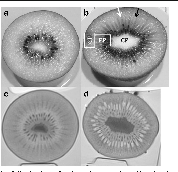
Figure 2 From Everyman S Prostate Phantom Kiwi Fruit Substitute For Human Prostates At Magnetic Resonance Imaging Diffusion Weighted Imaging And Magnetic Resonance Spectroscopy Semantic Scholar from d3i71xaburhd42.cloudfront.net May promote healthy skin and hair: Deliciosa in 1984 and reserved the name a. Kiwi fruit known as chinese gooseberry is a rich source of nutrients like antioxidants, phytonutrient.
The kiwifruit is a small fruit approximately 3 inches long and weighing about four ounces.
It comes with the oval shape and about the size of a large hen's egg. This family of kiwis includes the variety you're probably most used to eating, being about the size of a large egg with a fuzzy choose fragrant, slightly soft fruits. Once you know how to determine whether a kiwi is ripe, this delicious green (or golden) fruit is easy to peel and prepare. The kiwifruit is a small fruit approximately 3 inches long and weighing about four ounces.
42+ Ct Anatomy Of Neck Spaces . Posted by radiologypics ⋅ march 21, 2013 ⋅ 1 comment. Atlas of the anatomy of the head and neck on a ct in axial, coronal, and sagittal sections, and 3d images. Neck Spaces 1 Parapharyngeal Space 2 Masticator Space 3 Carotid Space 4 Parotid Space 5 Mucosal Space 6 Perivertebral Radiology Imaging Radiology Anatomy from i.pinimg.com The 5 anatomical spaces of the infrahyoid neck. They are only created by pathology, e.g. 875 x 507 jpeg 87 кб. The anatomy of the neck is complex and, as a result, not very well understood. Deep neck space infections michael d. Ct anatomy of neck spaces rv. Learn vocabulary, terms and more with flashcards, games and other study tools. Fasciae & spaces of the neck. Source: pubs.rsna.org They are only created by pathology, e.g. Source: i2.wp.com Neck nod...
11+ Human Anatomy Abdomen Muscles . Next to it on both sides of the body is the internal oblique. The posterior abdominal wall is found medial to the. Core Stability Abdominals Back Pain Pouch Weak Core Priority Physical Therapy Llc from images.squarespace-cdn.com An interactive demonstration of the transversus abdominis muscle (insertion, origin, actions & innervations) featuring the iconic gbs illustrations. You can locate them by putting your the external oblique muscles allow flexion of the spine, rotation of the torso, sideways bending and compression of the abdomen. The posterior abdominal wall is found medial to the. Actions.—when the pelvis and thorax are fixed, the abdominal muscles compress the abdominal viscera by constricting the cavity of the abdomen, in which action they are materially assisted by the. Attached to the bones of the skeletal abdomen and lower back. Att...
13+ Skull Base Bone Anatomy Ct . It supports the structures of the face and provides a protective cavity for the brain. Given that the file is large, loading may take a few minutes. Middle Skull Base Plastic Surgery Key from plasticsurgerykey.com Working knowledge of the normal and variant anatomy of the skull base is essential for effective surgical treatment of disease in this area. The skull is a bone structure that forms the head in vertebrates. The maxilla, nasal bone, lacrimal bone, zygomatic bone, etc. Given that the file is large, loading may take a few minutes. The maxilla, nasal bone, lacrimal bone, zygomatic bone, etc. An overview of the bones of the skull, including the frontal, parietal and occipital bones. 2.1 skull base anatomy—anterior and middle skull base. This anatomic region is complex and poses surgical challenges for otolaryngologists and neurosurgeons alike. ...


Comments
Post a Comment