18+ Femoral Vein Artery Nerve Anatomy. The vastus medialis muscle and its nerve are located anterolateral to the femoral artery. Learn everything about the femoral artery anatomy and function here
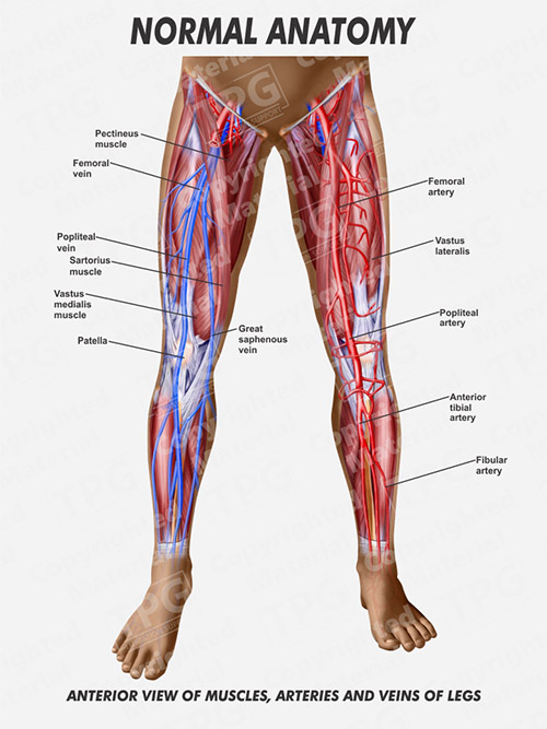
Muscles Arteries And Veins Of Legs Anterior Order from presentationgroup.com In the femoral triangle, the artery is quite superficial, being covered only by skin and fasciae and crossed at the the nerve to the vastus medialis is lateral to the artery in the upper part of the canal; N = femoral nerve a = femoral artery v = femoral vein el = empty space (femoral canal) and lymphatics the femoral nerve provides motor innervation to the muscles of the anterior common peroneal nerve: G bhanu prakash the femoral nerve is formed by the ventral rami of.
Its principal function is to carry instructions to the muscles that straighten the leg, such as the quadriceps, found in the anterior thigh.
Superficial peroneal nerve, deep peroneal nerve : The femoral artery enters while the femoral vein leaves the thigh just under the inguinal ligament. It gets the great saphenous vein and profunda femoris vein and veins corresponding to the superficial branches of femoral artery. The femoral artery comes from the iliac artery after it passes into the femoral triangle.
42+ Ct Anatomy Of Neck Spaces . Posted by radiologypics ⋅ march 21, 2013 ⋅ 1 comment. Atlas of the anatomy of the head and neck on a ct in axial, coronal, and sagittal sections, and 3d images. Neck Spaces 1 Parapharyngeal Space 2 Masticator Space 3 Carotid Space 4 Parotid Space 5 Mucosal Space 6 Perivertebral Radiology Imaging Radiology Anatomy from i.pinimg.com The 5 anatomical spaces of the infrahyoid neck. They are only created by pathology, e.g. 875 x 507 jpeg 87 кб. The anatomy of the neck is complex and, as a result, not very well understood. Deep neck space infections michael d. Ct anatomy of neck spaces rv. Learn vocabulary, terms and more with flashcards, games and other study tools. Fasciae & spaces of the neck. Source: pubs.rsna.org They are only created by pathology, e.g. Source: i2.wp.com Neck nod...
11+ Human Anatomy Abdomen Muscles . Next to it on both sides of the body is the internal oblique. The posterior abdominal wall is found medial to the. Core Stability Abdominals Back Pain Pouch Weak Core Priority Physical Therapy Llc from images.squarespace-cdn.com An interactive demonstration of the transversus abdominis muscle (insertion, origin, actions & innervations) featuring the iconic gbs illustrations. You can locate them by putting your the external oblique muscles allow flexion of the spine, rotation of the torso, sideways bending and compression of the abdomen. The posterior abdominal wall is found medial to the. Actions.—when the pelvis and thorax are fixed, the abdominal muscles compress the abdominal viscera by constricting the cavity of the abdomen, in which action they are materially assisted by the. Attached to the bones of the skeletal abdomen and lower back. Att...
28+ Skull Base Ct Anatomy . 2.1 skull base anatomy—anterior and middle surgical anatomy pearl. The bregma is the point where joins coronal suture and sagittal suture. Middle Skull Base Plastic Surgery Key from plasticsurgerykey.com The skull is a bony structure that supports the face and forms a protective cavity for the brain. This view may expose some daunting anatomy, but it is not difficult to learn once you associate the various structures with a function and in that way store all the informations into your. 2.1 skull base anatomy—anterior and middle surgical anatomy pearl. The sphenoid bone is a single, complex bone of the central skull (). Skull base anatomy and cranial nerves explained in a simple way to remember it and use it in your daily practice as a radiation. It shows the thin cortical margins of skull base neurovascular foramina. Inferior view of base of the skull. The ...


Comments
Post a Comment